The Visual Pathway
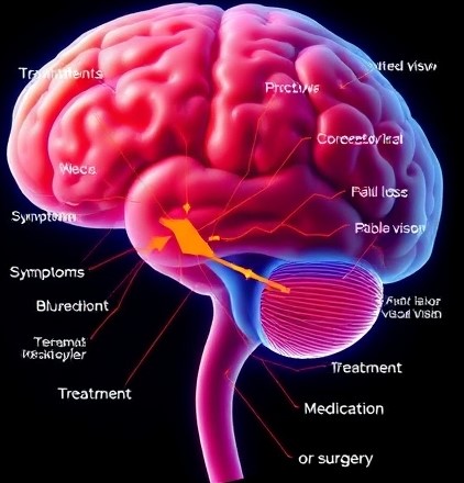
The Visual Pathway: At a Glance
Function: Transmits and processes visual signals for sight.
Symptoms: Blurred vision, field loss, or double vision.
Treatment: Therapy, medication, or surgery.

The Visual Pathway
The visual pathway is a complex system that allows us to process the visual information our eyes receive. While it may seem like the visual pathway begins at the cornea (where light first makes contact with the eye), the true pathway starts at the retina.
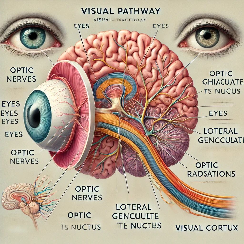
The following sections explain the key anatomical structures involved in the visual pathway, including the optic nerves, optic chiasm, optic tracts, lateral geniculate body, optic radiation, and the visual cortex
Anatomy of the Visual Pathway
While it may seem like the visual pathway begins at the cornea, where light first enters the eye, the actual visual pathway starts at the retina, where light is converted into electrical signals.
These signals then travel through a series of structures that relay, process, and interpret the visual information, ultimately allowing us to perceive the world around us. The main structures involved in the visual pathway are as follows:

Optic Nerves (CN II)
The optic nerves (Cranial Nerve II) are the first major structures in the visual pathway. They carry the electrical signals generated by the retina towards the brain. The signals from the retina’s ganglion cells (which are responsible for transmitting visual information) are bundled into the optic nerve fibers.
Each optic nerve transmits the information from one eye, so the left optic nerve transmits signals from the left visual field of both eyes, and the right optic nerve transmits signals from the right visual field.
Optic Chiasm
The optic chiasm is the point where the two optic nerves meet and partially cross. Here, some of the nerve fibers from each optic nerve cross over to the opposite side, while others remain on the same side.
This crossing of fibers is essential for binocular vision, as it allows visual information from the right side of both eyes to be processed by the left side of the brain, and visual information from the left side of both eyes to be processed by the right side of the brain. The crossing of these fibers is known as decussation

Optic Tracts
Once the nerve fibers have crossed over at the optic chiasm, they continue as the optic tracts. Each optic tract contains visual information from both eyes, but the information is organized by the visual field.
The right optic tract carries information from the right visual field of both eyes, while the left optic tract carries information from the left visual field. The optic tracts travel to the lateral geniculate nucleus (LGN) of the thalamus.
Lateral Geniculate Body (Lateral Geniculate Nucleus)
The lateral geniculate body (LGB), also known as the lateral geniculate nucleus (LGN), is a structure located in the thalamus, a part of the brain that acts as a relay station for sensory information. The
LGN is the primary processing center for visual signals. It processes basic visual information such as color, contrast, and brightness. After the visual information is processed, it is sent from the LGN to the visual cortex via a pathway called the optic radiation.
Optic Radiations
The optic radiations are a bundle of nerve fibers that carry visual information from the LGN to the visual cortex. These radiations spread out in the brain and ensure that the visual signals are directed to the appropriate areas of the cortex.
They transmit the information in such a way that the left optic radiation carries the visual information from the right visual field, and the right optic radiation carries the information from the left visual field.

Visual Cortex and Its Cortical Projections
The visual cortex is located in the occipital lobe at the back of the brain. It is the final processing area in the visual pathway where all the visual information is integrated. The primary visual cortex (V1) is responsible for processing basic visual features like orientation, color, and contrast.
The information then travels to higher-order areas of the cortex, such as the ventral stream, which processes object recognition and form, and the dorsal stream, which is involved in spatial awareness and movement. These projections help us recognize faces, read, navigate our environment, and more
How Visual Information Travels Through the Pathway
The journey of visual information begins when light enters the eye. The cornea and lens work together to focus light onto the retina, where photoreceptor cells (rods and cones) convert the light into electrical signals.
These signals are then transmitted through the optic nerve to the brain, where each structure in the visual pathway plays a critical role in processing and interpreting the information.
The Retina: The Starting Point of the Visual Pathway
The retina is the light-sensitive layer at the back of the eye, where photoreceptor cells (rods and cones) convert light into electrical signals. The signals are first processed by the ganglion cells and transmitted via their axons, forming the optic nerve.
The Optic Nerve: The First Link in the Pathway
The optic nerve is the first key link in the visual pathway, carrying electrical signals from the retina to the brain. It is made up of axons from the ganglion cells of the retina, transmitting visual information toward the brain.
The Optic Chiasm: Where the Pathways Cross
The optic chiasm is the crucial juncture where the optic nerves meet and partially cross. Here, fibers from each eye decussate (cross over) so that visual information from the left visual field of both eyes is processed by the right hemisphere of the brain, and visual information from the right visual field of both eyes is processed by the left hemisphere
The Lateral Geniculate Nucleus (LGN):
After passing through the optic chiasm, the visual information travels to the lateral geniculate nucleus (LGN) of the thalamus. The LGN acts as a relay station, processing basic visual information such as color, contrast, and brightness before sending it to the visual cortex for higher-level processing

The Optic Radiations: Connecting the LGN to the Visual Cortex
The optic radiations are nerve fibers that carry visual signals from the LGN to the visual cortex in the occipital lobe. These fibers help direct the information to specific areas of the cortex for detailed analysis of visual stimuli.
The Visual Cortex: Final Processing
The visual cortex is located in the occipital lobe, where visual information is processed into coherent images. The primary visual cortex (V1) handles basic visual features, and higher-order visual areas (in the ventral and dorsal streams) process more complex aspects, like object recognition and spatial awareness.
The Complexity and Precision of the Visual Pathway
The visual pathway is a highly organized and intricate system that ensures the efficient processing of visual stimuli. Each step of the pathway—from the retina to the visual cortex—plays a crucial role in transforming light into the images we interpret.
Any damage to these structures can lead to visual impairments, but understanding the pathway helps us appreciate the complexity of vision and aids in diagnosing and treating related disorders
