Ischemic Diabetic Retinopathy
Ischemic Diabetic Retinopathy (IDR) is a severe form of diabetic retinopathy characterized by extensive capillary non-perfusion within the retina. This condition leads to inadequate blood supply (ischemia),
which damages retinal tissues and stimulates the growth of abnormal blood vessels—a process known as neovascularization. Ischemic Diabetic Retinopathy (IDR) is considered a sight-threatening complication of diabetes and requires timely diagnosis and management to prevent irreversible vision loss
Ischemic Diabetic Retinopathy subtype of diabetic retinopathy is defined by significant areas of the retinal capillary network failing to supply adequate blood flow. The lack of oxygen delivery causes hypoxia, which is a driving factor for disease progression and complications
Signs and Symptoms
The signs and symptoms of Ischemic Diabetic Retinopathy IDR are as follows
- Reduced Visual Acuity: Vision becomes blurry or dim, especially in the affected areas.
- Floaters: Patients may notice spots or strings floating in their vision due to bleeding from new abnormal vessels.
- Dark Spots or Blind Spots: Resulting from areas of ischemia in the retina.
- Peripheral Vision Loss: The outer edges of vision may start to deteriorate before central vision is affected.
Causes
The causes of the Ischemic Diabetic Retinopathy are as
- Chronic Hyperglycemia: Prolonged high blood sugar damages retinal blood vessels.
- Hypertension: High blood pressure contributes to vascular stress.
- Dyslipidemia: Elevated lipid levels exacerbate vascular occlusion and damage.
- Duration of Diabetes: The longer a person has diabetes, the higher the risk of developing IDR

Features
The features of Ischemic Diabetic Retinopathy
- Microaneurysms: Tiny bulges in blood vessels that may rupture and leak fluid.
- Hemorrhages: Small or large retinal bleeds due to weakened vessel walls.
- Venous Beading: Enlarged and irregular veins indicative of ischemia.
- Cotton-Wool Spots: Microinfarcts in the retinal nerve fiber layer caused by ischemia.
- Capillary Dropout: Areas where the blood supply is completely absent
Diagnosis
The ways and techniques of the diagnosis of Ischemic Diabetic Retinopathy
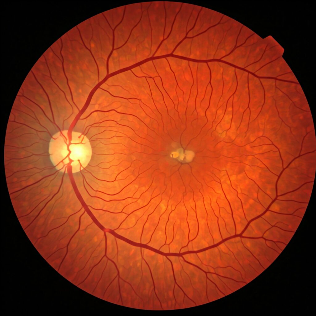
- Fundus Photography: High-resolution images of the retina reveal structural changes.
- Fluorescein Angiography (FA): This test uses a dye to highlight capillary non-perfusion and leakage.
- Optical Coherence Tomography (OCT): Non-invasive imaging to assess retinal thickness and detect edema.
- Visual Field Testing: Helps determine the extent of peripheral vision loss

Leads to Ischemia
Pathophysiology
Ischemia, or reduced blood flow, triggers the retina to release vascular endothelial growth factor (VEGF). VEGF promotes the formation of new blood vessels in an attempt to compensate for the oxygen deficit. Unfortunately, these new vessels are fragile and prone to leaking or bleeding, which can lead to complications like vitreous hemorrhage or tractional retinal detachment
Signs and Symptoms
- Neovascularization: Abnormal growth of blood vessels on the retina or optic disc.
- Fibrous Tissue Formation: Scar tissue accompanies neovascular growth, increasing the risk of retinal detachment.
- Vision Fluctuations: Vision may worsen suddenly due to hemorrhage or retinal damage
Causes
- Prolonged Ischemia: Persistent lack of oxygen triggers VEGF production.
- Retinal Hypoxia: Starvation of oxygen in the retinal tissues worsens disease progression
Features
- Proliferative Changes: Neovascular vessels extend beyond the retinal surface.
- Vitreous Hemorrhage: Blood leakage into the vitreous cavity obstructs vision.
- Retinal Detachment: Fibrovascular tissue pulls on the retina, detaching it from its base
Diagnosis
- Wide-Field Fluorescein Angiography: Maps the areas of capillary dropout and new vessel formation.
- Ultrasound Imaging: Detects vitreous hemorrhage or retinal detachment when the view is obstructed.
- OCT Angiography: Provides a detailed map of blood flow within the retina
Treatment
- Anti-VEGF Injections: Medications like ranibizumab or aflibercept block VEGF, reducing neovascular growth and leakage.
- Panretinal Photocoagulation (PRP): Laser therapy targets ischemic areas to reduce VEGF production.
- Vitrectomy Surgery: Removes blood-filled vitreous or scar tissue to restore vision.
- Glycemic Control: Maintaining optimal blood sugar levels prevents further ischemic damage.