Diabetic Retinopathy
At a glance: Diabetic Retinopathy
Early Symptoms: None
Later Symptoms: Blurry imaginative and prescient, floating spots in your imaginative and prescient, blindness
Diagnosis: Dilated eye exam
Treatment: Injections, laser treatment, surgical operation

Definition Of Diabetic Retinopathy
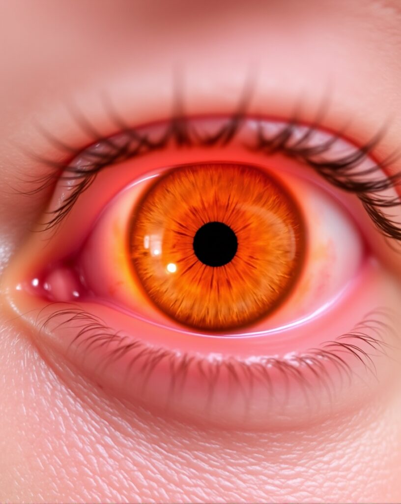
Diabetic retinopathy is one of the microvascular complications resulting from diabetes disease that affects retina as a result of the damage of the retina,
which is the back tissue in the eye that is responsible for light sensitivity. The high sugar levels in the blood are the major cause of the diabetic retinopathy.
This leads to the leakage of the blood vessels in the retina, swelling, and even jamming, which then leads to the growth of abnormal blood vessels.
It is divided into two main types:
Non-proliferative diabetic retinopathy (NPDR): The first step leading to that the blood vessels of the retina weaken of course the blood vessels do not grow abnormally.
Proliferative diabetic retinopathy (PDR): An advanced level characterized by the growth of abnormal blood vessels and the vitreous body. They can also cause severe problems with vision and blindness.

Causes Of Diabetic Retinopathy
One of the causes of the retina’s damage is the longevity of hypertensive hyperglycemia.
The increase of glucose in the body’s blood vessels can create pressure that can make the small blood vessels burst and be subjected to swelling, leakage, or be totally blocked.
These changes over time build up so that the blood circulation in the retina becomes impaired and the deficiency of oxygen caused by
it (ischemia) is added, which is a precipitating factor for the abnormal, weak growth of blood vessels (proliferation of neovasculature).
Other factors include:
Hypertension: Uncontrolled blood pressure can bring about the blood vessel damage.
Hyperlipidemia: The increase of cholesterol levels in the body can result in the formation of plaques on blood vessels,
which will end up being the main cause of atherosclerosis.
Inflammatory factors: The presence of type 2 diabetes can cause chronic retinal inflammation, which will damage the retinal blood vessels.
Risk Factor Of Diabetic Retinopathy
Several factors put a person at not so low odds of losing his vision from Diabetic Retinopathy. These are:
Duration of Diabetes: Diabetes that remains untreated over a prolonged period of time gives a person a higher than average risk for diabetes complications.
Type 1 or Type 2 Diabetes: Both of these types of diabetes are characterized by insulin resistance,
but the type 1 type is particularly affecting younger patients and throughout the duration of life, predominantly.
Poor Glycemic Control: Not effectively maintained sugar levels contribute to the increased risk very much.
Hypertension (High Blood Pressure): Unregulated blood pressure is also said to promote the early onset of retinal damage.
High Cholesterol Levels: Excessive cholesterol in the blood can lead to the injury of blood vessels.
Pregnancy: Diabetes primarily during pregnancy may accelerate or exacerbate DR in affected women.
Signs and Symptoms of Diabetic Retinopathy
Initially, DR often remains asymptomatic, say that is, symptoms are not noticed; therefore,
the doctor will need to continue regular eye exams for a diabetic patient. As the disease moves forward, the symptoms may include:
Blurry Vision: This is caused by the retina swelling.
Floaters: Tiny spots or strings that float in the visual field may indicate bleeding from abnormal blood vessels.
Dark or Empty Areas in Vision: Sometimes the ischemic part of the retina leads to some gaps in vision.
Fluctuating Vision: Blood sugar can affect vision clarity for a short while.
Impaired Color Vision: Damage of retinal causes a person to have a difficult time to distinguish colors.
Sudden Vision Loss: In the final stages, retinal detachment or bleeding of a large amount can cause the person to suddenly lose their vision.
Diagnosis Of Diabetic Retinopathy
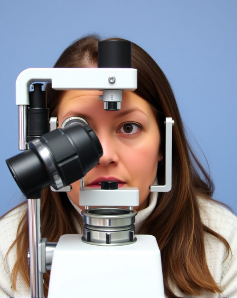
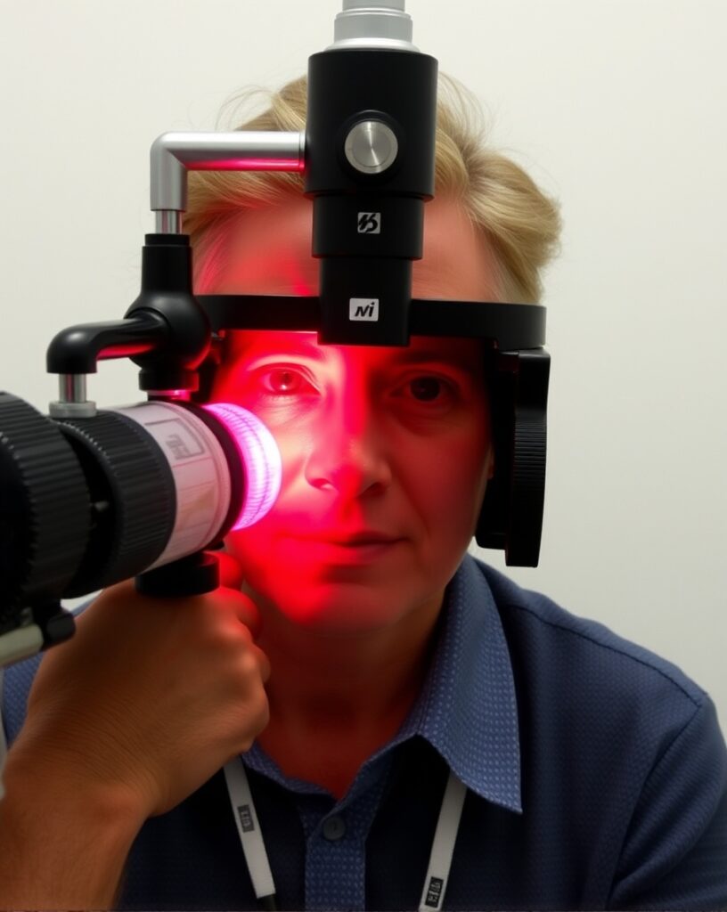
To diagnose Diabetic Retinopathy, uses a special light, called a retinoscope to look into the back of the eyes, through the small pupil.
Visual Acuity Test: Measures eyesight by having the patient read letters or numbers from a standardized chart.
Dilated Eye Exam: Dilating drops are instilled to open the eye’s pupil.
This allows the doctor to see the back of the eye and watch for abnormal blood vessel growth or new retinal problems

Optical Coherence Tomography (OCT): Using the optimal coherence tomography procedure,
clinicians can obtain and analyze high-resolution images of the retina with a view to detect the potential existence of edema or fluid buildup.
Fluorescein Angiography: A dye is injected into a vein, and a camera takes images as the dye travels through retinal blood vessels to reveal any leaks or blockages.
Prevention Of Diabetic Retinopathy
DR cannot be prevented in every case, but better diabetes control and a healthy lifestyle can reduce the likelihood of DR developing being detected:
Blood Sugar Control: Controlling blood glucose levels is essential in preventing diabetic retinopathy.
Keep regular Eye Exams: Regular annual eye exams will catch early signs of DR.
Blood Pressure Control: Keeping BP < 140/90 mmHg can help prevent DR or decrease its progression.
Cholesterol: Elevated levels of cholesterol can harm blood vessels and avoiding high-cholesterol foods lowers your risk.
Lifestyle Factors: Eating a balanced diet, exercising regularly and not smoking can keep you healthier and reduce the risk of DR.
Treatment Of Diabetic Retinopathy
Early Stages of DR
If your patient has mild NPDR, you may not need to treat right away but close monitoring is key.
Glycemic control: Better glycemic status can prevent progression of mild NPDR to severe DR.
Advanced Stages of DR
Laser Treatment (Pan-Retinal Photocoagulation):
This is a course of therapy that we do for many PDR, This includes vaporizing abnormal blood vessels or reviling leaky ones with a laser.
To control more blood leakage to prevent further retinal detachment.
Focal/Grid Laser Specific area with leaky vessels
Panretinal Photocoagulation: for PDR in a larger area of the eye to destroy abnormal vessels.
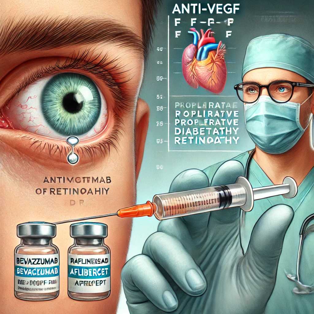
Anti-VEGF Agents (Injections)
Anti-VEGF drugs, including bevacizumab, ranibizumab and aflibercept prevent the activity of a protein called vascular endothelial growth factor (VEGF)
that stimulates abnormal blood vessel growth. Injections of anti-VEGF drugs are used to decrease leakage and retard progression in some cases with PDR.
These shots go straight into the eye and are generally repeated multiple times.

Steroid Injections:
An injection of corticosteroids into the eye may also be used to help reduce swelling in a complication called diabetic macular edema (DME) area related with DR.
