External Eye Structures
The external eye structures play a vital role in protecting and supporting vision. These components work in harmony to maintain eye health, guard against environmental hazards, and facilitate optimal vision. In this guide, we’ll explore each structure in detail, highlighting its anatomy, function, and common disorders.
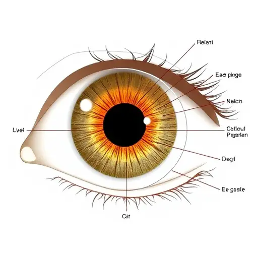
The external eye structures include the eyelids, eyelashes, conjunctiva, cornea, sclera, tear film, lacrimal apparatus, caruncle, and plica semilunaris.
Each of these components serves specific purposes, such as shielding the eye from injury, maintaining hydration, and ensuring clear vision. Together, they form the first line of defense against external threats and play a crucial role in overall ocular health.
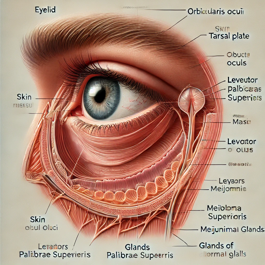
Eyelids
Anatomy of the Eyelids
The eyelids are thin, movable folds of skin and muscle. They consist of an upper and lower lid, supported by connective tissue called the tarsal plate. Internally, the eyelids are lined by the palpebral conjunctiva, which helps spread tears across the eye’s surface.
Function: Protection and Lubrication
The primary function of the eyelids is to protect the eyes from debris, excessive light, and physical injury. Blinking helps distribute the tear film, keeping the eye hydrated and free of irritants.
Common Eyelid Disorders
- Blepharitis: Inflammation of the eyelid margins caused by bacterial infections or blocked oil glands.
- Ptosis: Drooping of the upper eyelid due to weakened muscles.
- Stye: A painful lump caused by infected oil glands.
Eyelashes
Structure of Eyelashes
Eyelashes are rows of thick, curved hairs located at the edge of the eyelids. They are embedded in hair follicles connected to oil glands, which keep them lubricated.
Role in Eye Defense Mechanism
Eyelashes act as a barrier, preventing dust, dirt, and other particles from entering the eye. Their sensitivity triggers a reflexive blink, offering further protection.
Disorders of the Eyelashes
- Trichiasis: Abnormal inward growth of eyelashes, causing irritation and potential damage to the cornea.
- Madarosis: Loss of eyelashes due to conditions like alopecia or infections.
Conjunctiva
Layers of the Conjunctiva
The conjunctiva is a transparent membrane covering the sclera (bulbar conjunctiva) and the inner eyelids (palpebral conjunctiva).
Function: Lubrication and Immunity
This structure produces mucus and tears to keep the eye moist and comfortable. It also acts as a barrier against pathogens, thanks to its immune cells.
Conjunctival Diseases
- Conjunctivitis: Commonly known as pink eye, it causes redness and swelling due to infections or allergies.
- Pinguecula: A yellowish growth caused by UV exposure and dryness
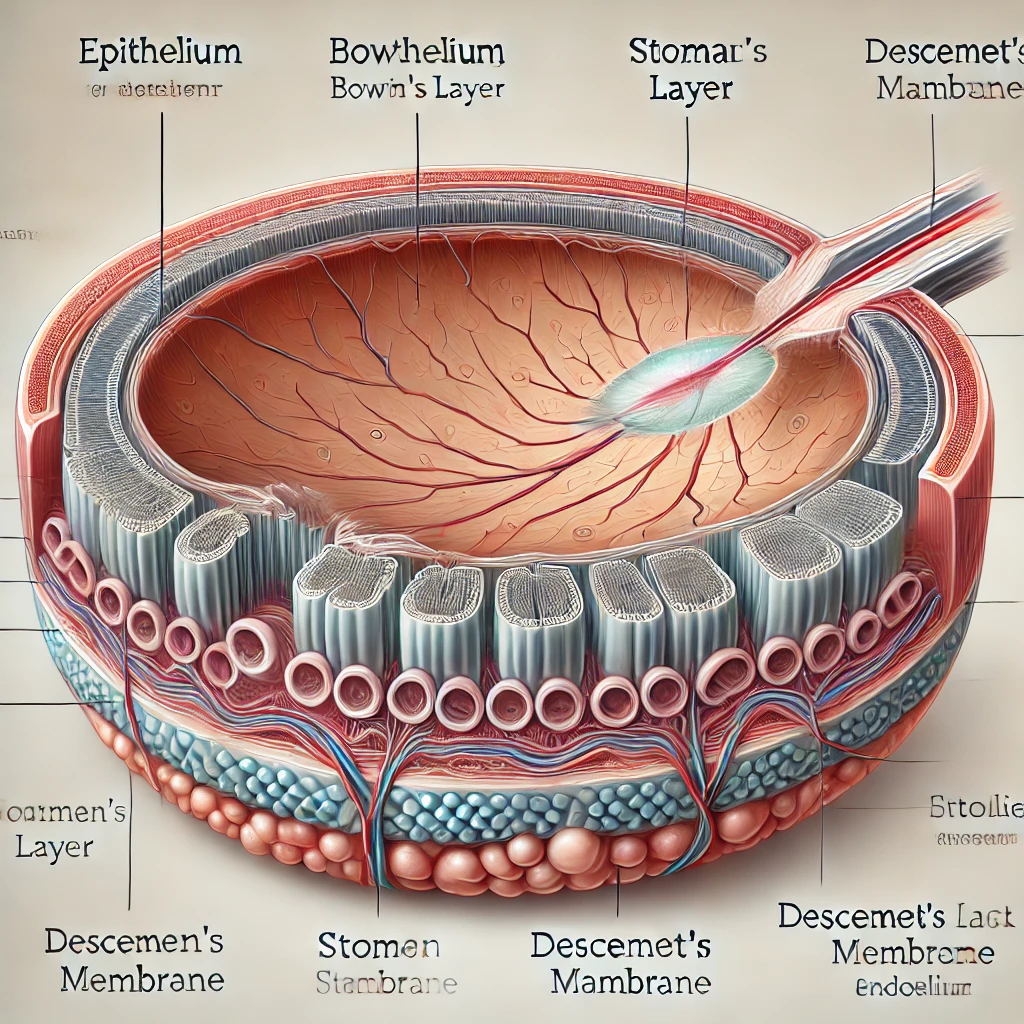
Cornea
Anatomy of the Cornea: Layers Explained
The cornea is the clear, dome-shaped outermost layer of the eye. It consists of five layers
- Epithelium: The outermost layer that regenerates quickly.
- Bowman’s Layer: A protective, tough membrane.
- Stroma: The thickest layer, providing strength and elasticity.
- Descemet’s Membrane: A thin, resilient layer.
- Endothelium: Maintains corneal clarity by regulating fluid levels.
Function: Transparency and Refraction
The cornea focuses light onto the retina, contributing to about 65-75% of the eye’s total refractive power
Common Corneal Disorders
- Keratitis: Inflammation caused by infections or injury.
- Corneal Ulcer: An open sore resulting from infections or trauma
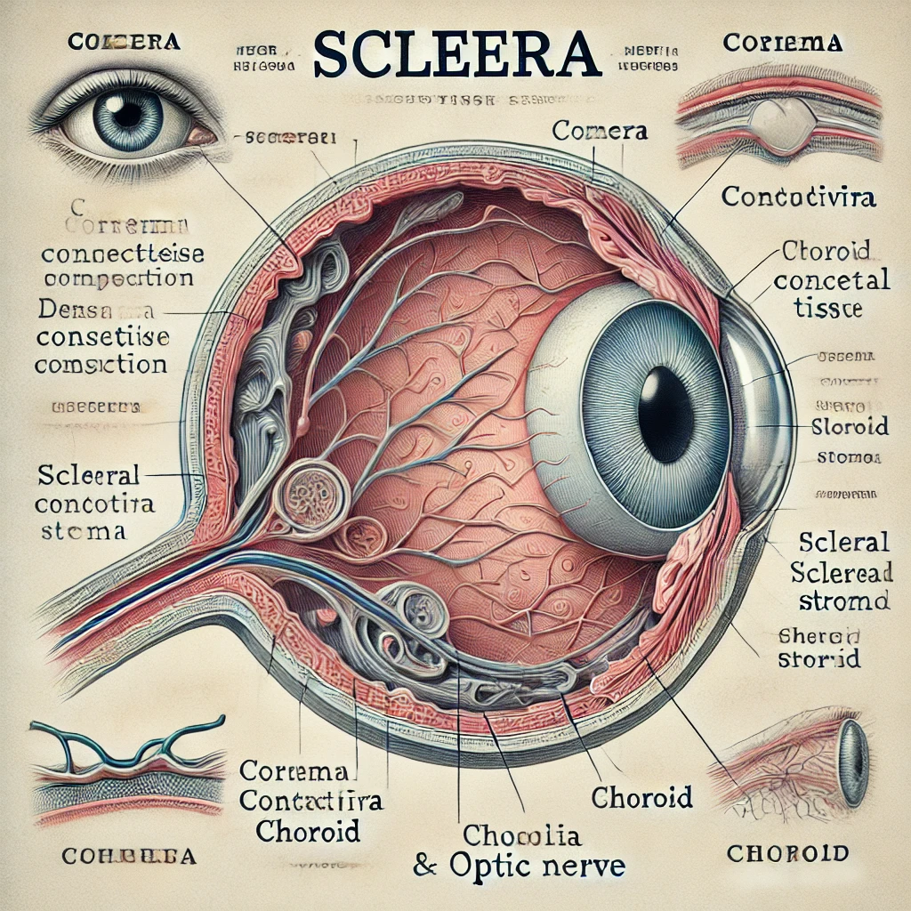
Sclera
Structure and Composition of the Sclera
The sclera, or the “white” of the eye, is made up of dense connective tissue. It provides structural integrity and protects the inner components of the eye.
Structure and Composition of the Sclera
The sclera, or the “white” of the eye, is made up of dense connective tissue. It provides structural integrity and protects the inner components of the eye.
Protective Role of the Sclera
By serving as a tough outer coating, the sclera resists injury and helps maintain the shape of the eyeball.
Protective Role of the Sclera
By serving as a tough outer coating, the sclera resists injury and helps maintain the shape of the eyeball.
Scleral Conditions
Scleritis: Painful inflammation often linked to autoimmune diseases
Tear Film and Lacrimal Apparatus
Composition and Layers of the Tear Film
The tear film has three layers:
- Lipid Layer: Prevents evaporation.
- Aqueous Layer: Provides moisture and nutrients.
- Mucin Layer: Helps tears spread evenly across the cornea.
Role in Maintaining Ocular Surface Health
The tear film keeps the eye hydrated, nourished, and free from irritants. It also facilitates smooth movement of the eyelids over the cornea.
Lacrimal Gland Anatomy and Tear Drainage System
The lacrimal gland, located above the outer corner of the eye, produces tears. Excess tears drain through the lacrimal puncta into the nasal cavity.
Caruncle and Plica Semilunaris
Understanding the Lesser-Known Structures
The caruncle is the small, fleshy area at the inner corner of the eye. The plica semilunaris is a crescent-shaped fold of conjunctiva adjacent to it.
Their Function in Tear Flow and Eye Movement
These structures assist in tear distribution and eye movement. The plica semilunaris is a remnant of a nictitating membrane (third eyelid) found in some animals.
Common Disorders of External Eye Structures
Overview of Diseases Affecting the Outer Eye
Disorders like blepharitis, conjunctivitis, and keratitis often impact the external eye structures, causing discomfort and vision problems.
Importance of Timely Diagnosis
Early detection and treatment can prevent complications and ensure better outcomes for eye health.
How to Maintain Healthy External Eye Structures
To keep your external eye structures in optimal condition, consider the following tips:
Tips for Hygiene and Care
- Wash hands before touching your eyes.
- Avoid rubbing your eyes to prevent infections.
- Clean your eyelids regularly, especially if prone to blepharitis
Preventing Environmental Damage
- Wear sunglasses to protect against UV rays.
- Use protective eyewear in dusty or hazardous environments.
- Stay hydrated and use artificial tears in dry conditions.