Diabetic Macular Edema (DME)
Diabetic Macular Edema (DME) is a significant complication of diabetic retinopathy (DR), which can develop at any stage of the disease, whether non-proliferative diabetic retinopathy (NPDR) or proliferative diabetic retinopathy (PDR).
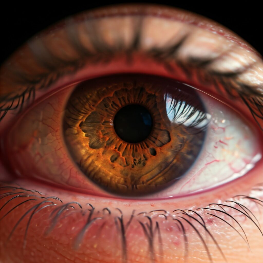
Diabetic Macular Edema (DME) occurs when damaged blood vessels in the retina leak fluid into the macula—the central part of the retina responsible for sharp, detailed vision. This fluid accumulation causes swelling in the macula, leading to visual distortion and potential vision loss. It is one of the leading causes of blindness among individuals with diabetes.
Signs and Symptoms
Diabetic Macular Edema (DME) does not always present noticeable symptoms in its early stages, which makes regular eye exams critical for individuals with diabetes. When symptoms do occur, they may include:
- Blurred or distorted central vision.
- Difficulty reading or recognizing faces.
- Colors appearing washed out or less vibrant.
- The appearance of dark or empty spots in the central vision.
- Increased sensitivity to light.
Since these symptoms can overlap with other eye conditions, professional diagnosis is essential to confirm DME.
Causes
- The primary cause of Diabetic Macular Edema (DME) is prolonged damage to the retinal blood vessels due to high blood sugar levels. Additional factors that can contribute to its development include:
- Chronic Hyperglycemia: Elevated blood sugar weakens the walls of retinal blood vessels, making them more prone to leaking.
- Hypertension: High blood pressure increases the risk of blood vessel damage in the eyes.
- Dyslipidemia: Abnormal cholesterol levels can exacerbate blood vessel problems.
- Duration of Diabetes: The longer a person has diabetes, the greater the risk of developing DME.
- Other Factors: Smoking and kidney disease can also increase the likelihood of DME
Features
Diabetic Macular Edema (DME) can be classified into two main types based on the pattern of fluid leakage and swelling in the macula:
- Focal DME:
- Characterized by localized leakage from specific points in the retina, often caused by microaneurysms (tiny bulges in blood vessels).
- Results in distinct, limited areas of swelling.
Typically easier to treat with targeted therapies
- Diffuse DME:
- Involves widespread leakage and swelling throughout the macula.
- Caused by extensive damage to retinal capillaries.
- Often more challenging to manage and may require combination treatments.
Understanding these subcategories is crucial for determining the appropriate treatment plan
Diagnosis
Diagnosing Diabetic Macular Edema (DME) involves a comprehensive eye examination conducted by an ophthalmologist or optometrist. Key diagnostic methods include:
- Visual Acuity Test: Measures the clarity of central vision.
- Dilated Eye Exam: Provides a detailed view of the retina and macula for signs of fluid accumulation.
- Optical Coherence Tomography (OCT): Uses light waves to create high-resolution images of the retina, allowing precise measurement of macular thickness and detection of swelling.
- Fluorescein Angiography: Involves injecting a dye into the bloodstream to highlight areas of blood vessel leakage and retinal damage.
- Fundus Photography: Captures detailed images of the retina to document the progression of DME.
These diagnostic tools are essential for confirming DME and tailoring treatment plans
Treatment
The management of Diabetic Macular Edema (DME) focuses on reducing macular swelling, preserving vision, and preventing further damage. Treatment options include
- Medical Management:
- Blood Sugar Control: Tight regulation of blood glucose levels can slow disease progression.
Blood Pressure and Cholesterol Management: Reducing these risk factors helps protect retinal health
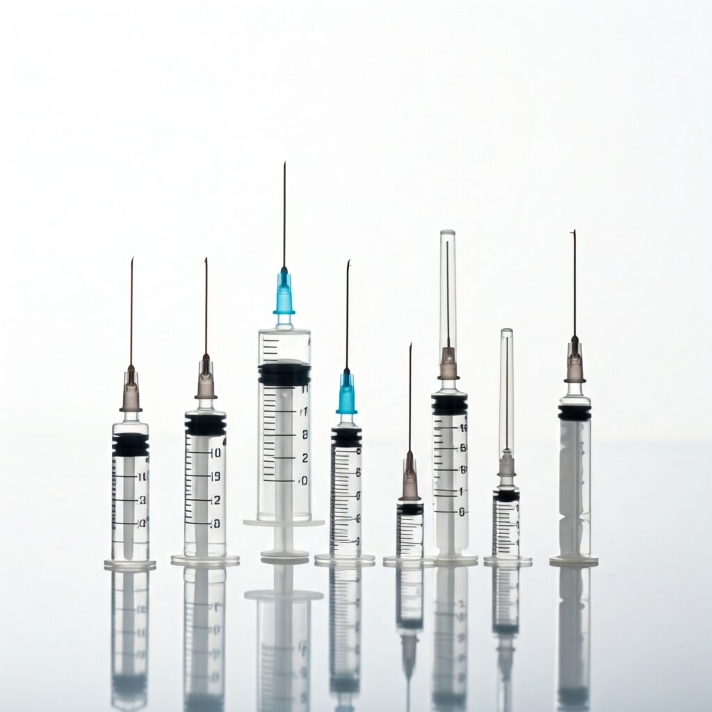
- Medications:
- Anti-VEGF Injections: These medications, such as ranibizumab, aflibercept, or bevacizumab, block vascular endothelial growth factor (VEGF), a protein that promotes abnormal blood vessel growth and leakage. Anti-VEGF therapy is a cornerstone in DME treatment.
- Steroid Injections or Implants: Used to reduce inflammation and swelling, particularly in cases unresponsive to anti-VEGF therapy.
- Laser Therapy:
Focal or Grid Laser Photocoagulation: Aimed at sealing leaking blood vessels to reduce fluid accumulation. It is particularly effective for focal DME
- Surgical Interventions:
Vitrectomy: May be performed in severe cases where the vitreous gel of the eye is pulling on the macula, contributing to swelling
- Lifestyle Changes:
Adopting a healthy diet, exercising regularly, and quitting smoking can improve overall outcomes and reduce complications